 17 Oct, 2026
Brain Aneurysm vs. AVM: Causes, Symptoms, Diagnosis and Treatment
Conditions affecting the blood vessels of the brain can sometimes develop without obvious warning signs. Brain aneurysms and arteriovenous malformations (AVMs) are two different types of cerebrovascular abnormalities that may require specialised medical evaluation.
Although both conditions involve abnormal blood vessels, they differ in their structure, development, potential complications, and treatment approaches. Understanding these differences can help patients recognise the importance of accurate diagnosis and individualised care.
What Is a Brain Aneurysm?
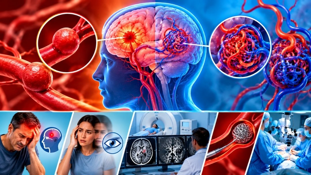
A brain aneurysm is a bulge that develops in a weakened area of a blood vessel wall. It can vary in size and shape and may occur in different arteries supplying the brain.
Some aneurysms remain stable and cause no symptoms, while others may grow or rupture. A ruptured aneurysm can cause bleeding around the brain, known as subarachnoid haemorrhage, and requires emergency treatment.
Possible symptoms of an unruptured aneurysm include pain around the eye, visual disturbances, or other symptoms caused by pressure on nearby structures. Many aneurysms, however, produce no noticeable symptoms before they are detected.
What Is an Arteriovenous Malformation (AVM)?
An arteriovenous malformation is an abnormal connection between arteries and veins that bypasses the normal capillary network.
Under normal circumstances, blood flows from arteries through tiny capillaries before entering veins. In an AVM, blood moves directly from arteries into veins through an abnormal network of vessels.
This altered blood flow can place stress on the affected vessels. Some AVMs are discovered incidentally, while others are identified after a seizure, headache, neurological symptoms, or bleeding.
The risks associated with an AVM depend on factors such as its size, location, vascular structure, and previous bleeding history.
Brain Aneurysm vs. AVM: Key Differences
Brain aneurysm: A localised bulge in the wall of a blood vessel.
AVM: An abnormal network of blood vessels connecting arteries and veins without the usual capillary pathway.
Possible complications: An aneurysm may rupture and cause bleeding around the brain. An AVM may also bleed and can sometimes cause seizures or other neurological symptoms.
Diagnosis: Both conditions may require specialised brain and blood-vessel imaging.
Treatment: Options vary and may include observation, endovascular procedures, surgery, or other specialised treatments, depending on the individual condition.
Because the two abnormalities are structurally different, their treatment plans are not interchangeable.
Common Symptoms of Brain Vascular Abnormalities
Symptoms depend on the location, size, and characteristics of the abnormality.
Possible symptoms include:
-
Persistent or unusual headaches.
-
Seizures.
-
Visual disturbances.
-
Weakness or numbness.
-
Difficulties with balance or coordination.
-
Speech or other neurological changes.
These symptoms can result from many medical conditions and do not automatically indicate an aneurysm or AVM.
A sudden, extremely severe headache, loss of consciousness, seizure, or new neurological weakness requires immediate emergency medical assessment.
How Are Aneurysms and AVMs Diagnosed?
Accurate diagnosis helps specialists understand the structure of the abnormal blood vessels and assess possible treatment options.
CT Scan: A CT scan may be used to evaluate suspected bleeding or other acute brain abnormalities.
MRI: MRI provides detailed images of brain tissue and can help identify certain vascular abnormalities and their effects on surrounding structures.
CT Angiography (CTA): CTA uses contrast material to create images of blood vessels and may help identify aneurysms and AVMs.
MR Angiography (MRA): MRA provides information about blood vessels using magnetic resonance imaging techniques.
Digital Subtraction Angiography (DSA): This specialised investigation can provide detailed information about the blood vessels feeding an AVM or the anatomy of an aneurysm. It may be recommended when precise vascular mapping is necessary.
The investigations selected depend on the suspected condition and the patient's clinical circumstances.
Treatment Options for Brain Aneurysms
Not every aneurysm requires immediate intervention. Treatment decisions depend on the aneurysm's characteristics, estimated rupture risk, location, patient health, and other relevant factors.
Endovascular Coiling
A catheter is guided through the blood vessels to the aneurysm, where small coils are placed to reduce blood flow into the abnormal bulge.
Surgical Clipping
A surgeon places a clip across the neck of the aneurysm to prevent blood from entering it. This may be appropriate for selected aneurysms.
Flow Diversion
For certain aneurysms, a specialised stent-like device may be placed in the parent blood vessel to redirect blood flow away from the aneurysm. Its suitability depends on the anatomy and clinical circumstances.
Monitoring
Some unruptured aneurysms may be monitored with follow-up imaging and management of relevant risk factors.
Treatment Options for AVMs
AVM management requires careful assessment of the potential risks of intervention and the risks associated with leaving the abnormality untreated.
Endovascular Embolisation
A catheter is used to deliver a specialised material into selected parts of the abnormal vascular network. Embolisation may be used as part of a broader treatment plan or, in selected circumstances, as a treatment approach on its own.
Microsurgical Removal
For certain AVMs, surgery may be considered to remove the abnormal vascular network. The decision depends heavily on its size, location, accessibility, and relationship to important brain structures.
Stereotactic Radiosurgery
This technique delivers focused radiation to an AVM to encourage gradual closure of the abnormal blood vessels over time. It does not usually eliminate the AVM immediately, and follow-up imaging is necessary.
Observation and Follow-Up
In some cases, a specialist may recommend monitoring rather than immediate intervention. This decision requires individual assessment of the AVM's characteristics and the patient's clinical circumstances.
The Role of Endovascular Neurosurgery
Endovascular neurosurgery uses catheter-based techniques to access and treat selected abnormalities within the blood vessels of the brain.
These techniques can be relevant to both aneurysms and AVMs, but the procedures used for each condition may differ significantly.
A specialist evaluates the vascular anatomy, reviews imaging studies, and discusses the potential benefits and risks of available approaches. Depending on the case, care may involve a multidisciplinary team that includes neurosurgeons, neurointerventional specialists, neurologists, and other healthcare professionals.
Why Individualised Evaluation Is Important
Brain aneurysms and AVMs vary considerably between patients. Factors such as the location of the abnormality, previous bleeding, symptoms, age, overall health, and treatment risks can influence the recommended approach.
Patients should discuss the purpose of each investigation, the risks associated with observation or intervention, available treatment alternatives, and the need for follow-up care.
Dr. Vivek Vaid, Brain & Spine Neurosurgeon and Endovascular Neurosurgeon, has a professional focus on neurosurgical and neurovascular conditions. Patients with a diagnosed or suspected cerebrovascular abnormality should seek assessment from a qualified specialist.
Frequently Asked Questions
Can an aneurysm or AVM exist without symptoms?
Yes. Both conditions may remain undetected until imaging is performed for another reason or symptoms develop.
Are brain aneurysms and AVMs the same condition?
No. An aneurysm is a bulge in a blood vessel wall, whereas an AVM is an abnormal connection between arteries and veins.
Does every AVM require surgery?
No. Treatment depends on the AVM's characteristics, associated risks, and the patient's overall health. Observation and different treatment approaches may be considered.
Can endovascular treatment be used for both conditions?
Yes, selected aneurysms and AVMs can be treated using endovascular techniques. However, the procedure and suitability differ according to the specific abnormality.
Conclusion
Brain aneurysms and AVMs are distinct cerebrovascular conditions that require accurate diagnosis and careful treatment planning. Understanding their differences can help patients recognise why specialist evaluation and individualised care are important.
Dr. Vivek Vaid's neurosurgical and endovascular practice is relevant to the assessment of selected neurovascular conditions. Patients should consult a qualified specialist to understand their diagnosis, available treatment options, and appropriate follow-up.